


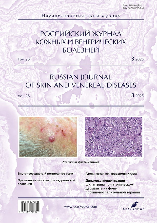
Атипичная фиброксантома кожи
- Авторы: Вертиева Е.Ю.1, Тертычный А.С.1, Цой Л.В.1, Хорошева Д.А.1, Бочарова Т.М.2
-
Учреждения:
- Первый Московский государственный медицинский университет имени И.М. Сеченова (Сеченовский Университет)
- Саратовский государственный медицинский университет имени В.И. Разумовского
- Выпуск: Том 28, № 3 (2025)
- Страницы: 233-239
- Раздел: ДЕРМАТООНКОЛОГИЯ
- URL: https://medbiosci.ru/1560-9588/article/view/313070
- DOI: https://doi.org/10.17816/dv677697
- EDN: https://elibrary.ru/MFHYSI
- ID: 313070
Цитировать

Аннотация
Атипичные фиброксантомы ― редкие кожные новообразования, относящиеся к группе фиброгистиоцитарных опухолей. Наиболее часто опухоль образуется в пожилом возрасте на фоне фотоповреждения кожи. Как правило, локализуется в области головы, плеч, верхней трети спины. Атипичная фиброксантома часто сочетается с другими опухолями кожи. Для неё характерны местнодеструктивный рост и крайне редкое метастазирование (1–2% случаев). Однако у пациентов на фоне иммуносупрессии возможно агрессивное поведение данной опухоли. Клиническая картина неспецифична. Опухоль представлена узлом или узелком розовой или красно-розовой окраски. Атипичная фиброксантома не обладает специфическими дерматоскопическими критериями. Характерны такие признаки, как полиморфные сосуды (линейные, точечные, клубочковые, древовидные) и хризалиды, которые не позволяют с уверенностью отличить её от клинически сходных новообразований только на основе осмотра. Схожесть атипичной фиброксантомы с такими агрессивными опухолями, как беспигментная меланома и карцинома Меркеля, представляет проблему для клинициста. Диагноз может быть достоверно установлен исключительно на основании характерных гистологических и иммуногистохимических признаков. Золотой стандарт лечения — хирургическое широкое иссечение опухоли. Возможно проведение операции по методу Моса. Другие методы терапии не рекомендованы в связи с высокой частотой рецидивов.
Мы представляем случай развития атипичной фиброксантомы у пациента в возрасте 72 лет на фоне фотоповреждения кожи в сочетании с другими опухолями кожи (базальноклеточные карциномы, которые пациент самостоятельно убирал жидким азотом). При гистологическом исследовании кожи выявлен инвазивный рост изъязвлённой полиморфноклеточной низкодифференцированной опухоли с выраженной анаплазией, участками эпителиоидно-клеточного, местами веретеновидно-клеточного строения. Дифференциальный диагноз проводился между меланомой, фибросаркомой, атипичной фиброксантомой. Пациенту проведено широкое иссечение опухоли, материал отправлен на гистологическое и иммуногистохимическое исследование. Послеоперационный период протекал без осложнений с формированием рубца; по результатам динамического наблюдения признаков рецидива опухоли не выявлено.
Полный текст
Открыть статью на сайте журналаОб авторах
Екатерина Юрьевна Вертиева
Первый Московский государственный медицинский университет имени И.М. Сеченова (Сеченовский Университет)
Автор, ответственный за переписку.
Email: ivertieva@gmail.com
ORCID iD: 0000-0002-1088-2911
SPIN-код: 3712-8453
канд. мед. наук
Россия, МоскваАлександр Семенович Тертычный
Первый Московский государственный медицинский университет имени И.М. Сеченова (Сеченовский Университет)
Email: atertychyy@gmail.com
ORCID iD: 0000-0001-5635-6100
SPIN-код: 5150-0535
д-р мед. наук, профессор
Россия, МоскваЛариса Валерьевна Цой
Первый Московский государственный медицинский университет имени И.М. Сеченова (Сеченовский Университет)
Email: dr.lvtsoy@gmail.com
ORCID iD: 0000-0001-9072-2311
SPIN-код: 9581-6228
канд. мед. наук, доцент
Россия, МоскваДиана Алексеевна Хорошева
Первый Московский государственный медицинский университет имени И.М. Сеченова (Сеченовский Университет)
Email: 5_97@inbox.ru
ORCID iD: 0009-0006-1296-5848
Россия, Москва
Татьяна Михайловна Бочарова
Саратовский государственный медицинский университет имени В.И. Разумовского
Email: tatuana.bocharova@yandex.ru
ORCID iD: 0009-0002-4361-1357
Россия, Саратов
Список литературы
- Rosenfeld D, Alam M, van Tine B, Council ML. Atypical fibroxanthoma: A malignant tumor of the skin and soft tissue. J Am Acad Dermatol. 2020;83(6):e429–e430 doi: 10.1016/j.jaad.2020.07.022
- Agaimy A. The many faces of atypical fibroxanthoma. Semin Diagn Pathol. 2023;40(4):306–312. doi: 10.1053/j.semdp.2023.06.001
- Ríos-Viñuela E, Pons Benavent M, Monteagudo C, et al. Atypical fibroxanthoma and pleomorphic dermal sarcoma: A two-center retrospective study of 74 cases. Actas Dermosifiliogr. 2022;113(6):654–656. doi: 10.1016/j.ad.2021.09.008
- Helwig EB, May D. Atypical fibroxanthoma of the skin with metastasis. Cancer. 1986;57(2):368–376. doi: 10.1002/1097-0142(19860115)57:2<368::aid-cncr2820570230>3.0.co;2-n
- Bitel A, Schönlebe J, Krönert C, Wollina U. Atypical fibroxanthoma: An analysis of 105 tumors. Dermatol Ther. 2020;33(6):e13962. doi: 10.1111/dth.13962
- Pitarch G. Dermoscopic rainbow pattern in atypical fibroxanthoma. Actas Dermosifiliogr. 2014;105(1):97–99. doi: 10.1016/j.ad.2012.11.010
- Alves R, Ocaña J, Vale E, et al. Basal cell carcinoma and atypical fibroxanthoma: An unusual collision tumor. J Am Acad Dermatol. 2010;63(3):e74–e76. doi: 10.1016/j.jaad.2009.11.685
- Fisher JC, Jones M, Hurd DS. Atypical fibroxanthoma: A case report and literature review. J Am Osteopath Coll Dermatol. 2017;37(2):17–19.
- Piras V, Ferreli C, Atzori L, et al. Atypical fibroxanthoma/pleomorphic dermal sarcoma of the scalp with aberrant expression of HMB-45: A pitfall in dermatopathology. Pathologica. 2020;112(2):105–109. doi: 10.32074/1591-951X-39-19
- Nguyen CM, Chong K, Cassarino D. Clear cell atypical fibroxanthoma: A case report and review of the literature. J Cutan Pathol. 2016;43(6):538–542. doi: 10.1111/cup.12696
Дополнительные файлы


